| Collection, Preservation and Archival Storage of Crustaceans
Compiled by Joel W. Martin Natural History Museum of Los Angeles County Last Updated: 12 December 2004 |
 |
Introduction
Crustaceans are the fourth most speciose group of metazoans on earth, behind insects, mollusks, and chelicerates, with some 65,000 described species and an unknown number still awaiting discovery and description. The group's morphological diversity (disparity) is even greater than its species diversity. The various classes of Crustacea (the exact number depending on which classification you follow) contain far more diversity, as well as more species, than all birds, fishes, reptiles, amphibians, and mammals combined (Martin and Davis, 2001). The group contains the familiar crabs, shrimps, and lobsters, but also a host of other and smaller groups such as fairy shrimp, seed shrimp, amphipods, isopods, copepods, barnacles, and many more. They are also ecologically diverse, occurring in a surprisingly large number of habitats and ecological niches (Martin and Davis, 2001). Not surprisingly, it is difficult to make generalizations for the collection, fixation, preservation and archival storage that would apply to all members of so diverse a group.
In general, crustaceans have in common the following features:
- An external skeleton (cuticle) that is composed of chitin and that may contain various minerals and salts;
- Two pairs of pre-oral appendages, usually termed antennules and antennae (sometimes called first and second antennae).
- A nauplius larval stage during development (although this has been bypassed in the development of many groups, especially among the malacostracans;
- A head region composed of 5 fused segments, each bearing an appendage. In most of the crustacean groups these appendages are referred to as the anatennule, antenna, mandible, maxillule, and maxilla. In many groups the first few thoracopods are also modified into feeding appendages; when this is the case, these appendages are referred to as maxillipeds.
Extreme modification of the basic "bauplan" above is the rule rather than the exception, to the point that the above features are almost never all present within a single taxon.
There are, depending on which classification you follow, approximately six major lineages or classes of extant crustaceans generally recognized, although there is considerable debate about the composition and monophyly of these lineages. The six classes -- Branchiopoda, Cephalocarida, Maxillopoda, Remipedia, Ostracoda, and Malacostraca -- differ greatly as to the diversity within each; some classes (e.g. the Malacostraca and the probably-non-monophyletic Maxillopoda) are far more diverse than others. Identification to class depends on the number of body somites, and their appendages, tagmatization, larval types, etc.
"Picture keying" is actually acceptable as a first cut for sorting to subclass or order, as most of the major crustacean groups can be recognized as shown below: On the other hand, there are notable cases of convergence within the Crustacea, such as the remarkably "crab-like" anomurans and the true crabs (brachyurans), and in most instances a specialist should be consulted for identification to family level or below. There are an estimated 855 currently recognized extant families (Martin and Davis, 2001, with updates), and it is not possible to provide diagnoses or descriptions of each family here.
Crustacean Life History Stages
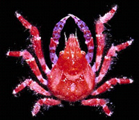
 Most crustaceans exhibit metamorphic development, and larval forms are numerous and diverse. Indeed, early workers who believed that some larval forms were adults named them, such that there is an entire vocabulary of crustacean larval types and descriptive terms. Collecting crustacean larvae is most often accomplished using plankton nets towed behind ships at sea. Rearing larvae from ovigerous adults is also commonly employed, and this method has resulted in most of our descriptions of "known" larvae (i.e., studies where the larval form and the adult are known to belong to the same species).
2. Documentation
Whenever possible, the act of collecting and/or observing crustaceans should be accompanied by thorough documentation. Documentation can take many forms, and not every form of documentation is desirable or even possible for each collecting event. Below are some of the more common forms of documentation. (See also General Protocols).
Transects: Because of their mobility, crustaceans are not often the targets of traditional "repeated" transect studies, with the exception of barnacles and some of the larger habitat-specific decapods encountered in, or close to, certain specific habitats (e.g. spiny lobsters on reefs). General transect protocols can be seen in the General Protocols section.
Photography: Because crustaceans so easily lose their coloration when fixed and preserved, field workers are strongly advised to take note of, and preferably to document using digital color photography, any colors or patterns seen on the freshly-collected specimens. These colors and patterns have been shown to have taxonomic and phylogenetic information, and usually such patterns are irretrievably lost during or after preservation.
Specific methods for color photography of crustaceans can be found on the Guana Island Marine Invertebrate Survey web site, http://www.nhm.org/guana/bvi-invt/bvi-surv/photogra.htm. For video photography and obtaining still images from video see the general protocol.
Orientation of Specimen in the Photograph
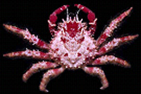 Crustaceans that are somewhat dorsally-ventrally flattened (e.g. most isopods, crabs, lobsters, etc.) can be posed and photographed from a dorsal perspective fairly easily (at right). Size determines whether the photographer should use a macro lens, dissecting microscope, or compound microscope. In nearly all cases, the best results are achieved by placing the specimen in clear, clean water to avoid matting of setae and glare reflecting off the cuticle, and to ensure a more natural appearance of the specimen; the photograph is taken through the water.
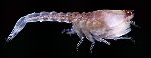
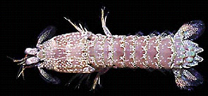
Taxa that are laterally compressed, such as most shrimp (below left), stomatopods (below right) and large amphipods, can be placed in a water-filled miniature, narrow-walled "aquarium" made
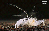
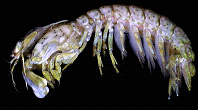
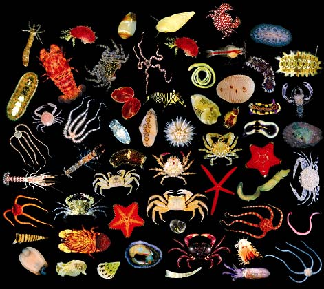
for that purpose out of small pieces of acrylic (see Guana Island Marine Invertebrate Survey web site). Although use of a neutral gray color background is sometimes advocated, we have obtained excellent results using a black background, which is facilitated by placing a piece of black felt or velvet beneath the specimen before taking the photograph. A solid black background also facilitates "merging" individual photographs into plates (see photograph below and the Photography methods section on the Guana Island Marine Invertebrate Survey web site).
 |
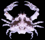 The angle of lighting can also be very important. Uniform lighting from above or from an angle of approximately 45° in general is better for overall color pattern, whereas indirect light or light from the side can accentuate three-dimensional features such as grooves and spines (e.g. at right).
Most photographs, regardless of their quality, are insufficient to allow identification of the subject to species level unless additional photographs, usually at higher magnifications, are also taken. Such additional taxonomic photographs should include close up photographs of the ventral surface and appendages (including, where possible, the mouthparts).
3. Collecting
Permits: Most collectors are now aware of the critical importance of obtaining proper permits for collecting any crustacean. Museums often are not allowed to accept or accession any specimens without proper permits and other documentation in hand to indicate the legality of the collecting expedition.
Some crustaceans are listed in the CITES Appendices. Collection of crustaceans from U.S. waters and subsequent transport within the U.S. does not require a CITES permit, but transport across international borders requires an export permit from the CITES Management Authority in the country of origin. Collections made in state waters (up to 3 miles offshore) need a state permit, and those in Federal waters need a Letter of Acknowledgement of scientific research, which is available from regional NMFS offices. To collect from Marine Reserves, you need special permission from the reserve management authority. These permits are granted by regional fisheries science centers. A list of these centers is available at http://www.noaa.gov/fisheries.html.
Collecting in foreign countries or waters is often greatly facilitated by working cooperatively with workers in that country; collecting under local national permits and subsequent long-term borrowing of materials is often the preferable, and sometimes the only, way to do this.
Collecting Methods
The known methods for collecting crustaceans are numerous and varied, although many of the more commonly employed methods are the same as those used for other marine invertebrates. These methods include (but are not limited to) hand-held nets, trawls, dredges, collecting via SCUBA diving (by hand or with nets or other devices), plankton net tows, ROV and manned submersible collecting devices, and many more. Traps (some baited, some using a light source as the attractor) are also commonly employed. Various pumps (e.g. "yabby" pumps) are used for tubicolous or fossorial species. Meiofaunal and other very small crustaceans often require other, more taxon-specific, methods.
Collection Documentation
A good "collection event" entails thorough documentation of the site and the habitat, collection of a specimen or several specimens, labeling of the specimen and some form of preservation.
4. Preservation
Fixatives and Preservatives
 Fortunately, despite their staggering morphological diversity, crustaceans are amenable to fixation and preservation using relatively few techniques, and they do not present insurmountable problems for preservation and long-term storage. As with most other groups of marine organisms, the choice as to which fixative and/or preservative to use depends ultimately on what the collection will be used for in the future.
It is important to note that the words fixation and preservation are used casually and often incorrectly in the collection care literature. There is a distinct difference between a fixative and a preservative. In general, fixation is more desirable for histological or detailed morphological studies, but specimens should never be stored in any fixative for long (or even short) periods of time, and indeed, with many crustaceans, fixation may not be necessary at all (as opposed to preservation in ethanol or other preservative).
The ever-present cuticle, varying among taxa in thickness, permeability, and incorporated salts and minerals, presents something of a barrier to fixatives and preservatives. For large crustaceans, especially among the Decapoda, it may be necessary to penetrate the cuticle by inserting a hypodermic needle beneath the rear of the carapace in order to inject some fixative or preservative into the large cephalothoracic region. This will prevent rotting of the tissues there, which otherwise might not be contacted by the fixative or preservative for several days.
Fixatives
A fixative actually "fixes" a specimen by stabilizing (cross-linking) the proteins within its tissues such that long afterwards, the tissues will still retain a semblance of their appearance in life. Additionally, fixing usually raises the refractive index of the tissue making it more susceptible to staining. By definition, then, a fixative is a toxic chemical with a strongly adverse affect on tissue, in a sense chemically "freezing" the tissues permanently in place. The most common fixative for crustaceans historically has been formalin. When purchased commercially (where it is usually labeled ("formaldehyde solution"), this is a 37%-40% aqueous solution of the gas formaldehyde. To make "10% formalin" (the most commonly used fixative), the commercial solution must be diluted using 9 parts water to 1 part commercial formaldehyde solution. For marine crustaceans, to prevent distortion due to osmotic forces, the 9 parts water should be clean, filtered seawater. The resulting solution is acidic and therefore is detrimental for specimens containing CaCO3, which unfortunately is present in the carapace of many crustaceans. To neutralize or "buffer" the acid content, small amounts of sodium hydroxide, sodium tetraborate, or the organic base hexamine are sometimes added. However, this buffering is apparently temporary, as buffered formalin will become acidic again some time after buffering; crustaceans should never be stored in formalin for extended periods of time.
Fixation should be limited to a period of a few hours to a few days -- long enough so that all tissues come in contact with the fixative but not so long that the crustacean becomes brittle, which can happen if left in formalin for too long a period. A 24-hour period is probably ideal. "Relaxing" the specimen (see below) by adding some kind of nacotizing agent (e.g. chloral hydrate, epsom salts, menthol, or other chemicals; see Knudsen, 1966: 510) to the water in which it is being held prior to killing and fixation or preservation is helpful for avoiding autotomy (see below). Softening agents such as propylene phenoxytol or (more commonly) propylene glycol or glycerol can be added in small amounts to maintain flexibility of the appendages, as in the use of Steedman's solution for planktonic crustaceans (Lincoln and Sheals, 1979). Following fixation, the specimen should be rinsed thoroughly in fresh or distilled water, after which it is usually transferred to 70% ethyl alcohol (ethanol) for long-term storage (see below).
Formalin denatures the DNA (or rather makes it unavailable by the binding of the surrounding histones) so that it is far more difficult, and in some cases impossible, to extract DNA for molecular studies in the future. Actual damage to the DNA might be caused not so much by the formalin as by the associated low pH (even, in some cases, after buffering, as the acidity apparently increases over time in stored collections; see Bucklin and Allen, 2003). Although there are reports of success in extracting DNA from formalin-preserved crustaceans, some even from archival collections decades old (e.g. France and Kochler, 1996; Bucklin and Allen, 2003; Schander and Halanych, 2003), if sequencing or other gene-based work is desired it would be best to avoid fixation altogether. The relatively recent trend toward "fixing" crustaceans in 95% ethyl alcohol, if they cannot be cryopreserved (frozen), assures their usefulness in DNA extraction (though not RNA or proteins) at least for the near future (e.g. Dessauer et al., 1996), although ethanol storage over long periods of time also complicates DNA extraction. Other studies of non-cryopreservation methods for "preserving" tissues of marine invertebrates for molecular work include Dawson et al. (1998; DMSO-NaCl, NaCl/CTAB, Fukatsu (1999; acetone), and Lee and Beynon (2004; potable alcohol and nail varnish remover), the last of which used crustaceans (balanoid barnacles) as the subject.
Other fixatives used for crustaceans include glutaraldehyde (mostly for SEM work), Davidson's solution, Bouin's solution (a mixture of picric acid, formaldehyde, and acetic acid) and osmium tetroxide is sometimes used as a "post fixative" for specimens destined for electron miscroscopy, as it is an extremely strong (and extremely toxic) fixative that stabilizes the cuticle. With all fixatives, extreme care must be used at all times, including the use of fume hoods, safety glasses, gloves, and proper ventilation.
Preservatives
A preservative, in contrast to a fixative, is a solution in which the specimen can be stored and maintained without further degradation. By far the most common preservative in regard to storage of crustaceans is ethyl alcohol, usually in a concentration of 70 to 75%. As with the fixatives, occasionally propylene glycol or glycerol are added in small amounts to maintain flexibility of the appendages and to slow down the dessication of the specimen, an ever-present problem in large collections of crustaceans. The primary role of the preservative is to provide an environment in which bacterial and other infestations and contaminations are unlikely, thus maintaining the specimens over long periods of time. For crustaceans, there is a "rule of thumb" suggesting that a ratio of at least 3:1 (3 parts ethanol to 1 part crustacean) is needed, possibly to keep the percentage of ethanol from becoming too low as fluids within the specimen's tissues seep out. Crustaceans placed directly into ethyl alcohol will last for years and can be used for some morphological work (such as illustrations of the entire specimen) and limited molecular work (see general discussion). Anecdotal accounts suggest that high concentrations of ethanol can cause the specimen to become brittle and less amenable to manipulation.
Because ethyl alcohol is the primary component of liquor, researchers can use, and often have used, commercially-purchased hard liquors (vodka, gin, tequila, etc.) in the appropriate strengths for temporary preservation of crustaceans. In our Caribbean study of shallow-water invertebrates (Zimmerman and Martin, 2004), we purchased and used cases of 150-proof (= 75% ethyl alcohol by volume) rum, which was less expensive by far than purchasing pure ethanol in the Caribbean and had the added benefit of creating a pleasing aura in our makeshift lab. Similar reports abound, and appear to be limited only by the taste (literally in some cases) of the investigator. There have been no studies on what effect, if any, other ingredients or additives in such drinks have on the long-term well being of the specimens (not to mention the researchers).
Relaxation of Specimens
Crustaceans, like some other arthropods, can throw off (autotomize) a limb when alarmed, and this behavior is often seen when the animal is exposed to a fixative or preservative. This behavior is seen mostly in the decapods and is believed to be an escape or predator-avoidance mechanism. "Relaxing" the specimen by adding some kind of nacotizing agent (e.g. clove oil, magnesium chlorate, chloral hydrate, epsom salts, CO2 gas, menthol, or other chemicals; see Knudsen, 1966: 510) to the water in which it is being held prior to fixation is helpful for avoiding autotomy. Limb autotomy also can be avoided by chilling the live specimen (immersing it in ice water for a few hours) and then transferring it to the desired fixative or preservative (especially for warm-water or terrestrial crustaceans). Chilling the specimen by immersing it in ice water has the additional benefit of maintaining the crustaceans' color, at least temporarily. Because the animal is freshly dead and can be posed easily, the period between immersion in ice water and fixing or perserving can be used for color photography. Relaxing a specimen prior to fixing or preserving it has other benefits as well, including better flexibility of the preserved specimen.
Softening agents such as propylene phenoxytol or (more commonly) propylene glycol or glycerol also can be added in small amounts to maintain flexibility of the appendages, as in the use of Steedman's solution for planktonic crustaceans (e.g. see Lincoln and Sheals, 1979). Addition of these chemicals to the preservative may have undesirable consequences for some future uses (e.g. SEM work).
Dried Specimens
Drying, as an intentional form of specimen preservation and storage, should be avoided for crustaceans. Although the resulting specimen may be suitable for some molecular work, it is essentially useless for any other purpose, and it will eventually fall apart.
5. Archival Storage
Whether the specimens are fixed or preserved initially, they should at some point be rinsed in water (if fixed) and transferred to 70 or 75% ethanol for long-term storage. Archival storage includes clear, clean 70 to 75% ethanol, archival-quality glass jars and lids (the best of which now come with polypropylene inserts in the lid), and archival-quality label paper with printed collecting and preservation data. The label is an often-overlooked but critical part of the collection, and it very often makes the difference between a valuable scientific specimen and a specimen that is useless for scientific purposes because there are no associated data (see Zala et al., in press, for a test of how different printing techniques last in different preservatives). Although different groups of crustaceans will vary with regard to the "fields" that are important (e.g., collection depth is not usually an important consideration when dealing with notostracans or terrestrial isopods), the label should a sufficient amount of clearly legible information.
Avoid at all costs the temptation to include labels bearing only the "station" or "field" number, believing that at some point a future worker will be able to extract the collecting data from your notebook, field journal, or spreadsheet. In Los Angeles, we have discarded tons (literally) of specimens, many of which came from enormously expensive studies that were conducted over a period of several years, because the associated data either were lost or were not provided with the collection. If the specimen is worth collecting, then it is also worth labeling correctly and informatively.
References
Bucklin, A., and L. D. Allen. 2003. MtDNA sequencing from zooplankton after long-term preservation in buffered formalin. Molecular Phylogenetics and Evolution30: 879-882.
Dawson, M. N., K. A. Rskoff, and D. K. Jacobs. 1998. Field preservation of marine invertebrate tissue for DNA analyses. Molecular Marine Biology and Biotechnology 7: 145-152.
Dessauer, H. C., C. J. Cole, and M. S. Hafner. 1996. Collection and storage of tissues. In: Hillis, D. M., et al. (editors), Molecular Systematics. Sunderland, Massachussetts; Sinauer.
France, S., and T. D. Kocher. 1996. DNA sequencing of formalin-fixed crustaceans from archival research collections. Molecular Marine Biology and Biotechnology 5: 304-313.
Harvey et al., 2001. Crustacea. In: Atlas of Invertebrate Larvae.
Knudsen, J. W. 1966. Biological Techniques: Collecting. Harper & Row, Publishers, New York.
Lee, P. L. M., and C. M. Beynon. 2004. Effectiveness of alternative organic solvents in field preservation of whole barnacles for PCR analyses. Journal of the Marine Biological Association of the United Kingdom 84: 747-749.
Lincoln, R. J., and J. G. Sheals. 1979. Invertebrate Animals: Collection and Preservation. British Museum (Natural History), Cambridge University Press.
Martin, J. W., and G. E. Davis. 2001. An updated classification of the Recent Crustacea. Natural History Museum of Los Angeles County, Science Series No. 39: 1-124.
Schander, C., and K. M. Halanych. 2003. DNA, PCR and formalinized animal tissue -- a short review and protocols. Organisms, Diversity & Evolution 3(3): 195-205.
Zala, K., N. D. Pentcheff, and R. Wetzer. In press. Laser-printed labels in wet collections: Are they holding up? Collection Forum.
Zimmerman, T. L. and J. W. Martin. 2004. Artificial reef matrix structures (ARMS): an inexpensive and effective method for collecting coral reef-associated invertebrates. Gulf and Caribbean Research 16: 59-64.
Acknowledgments: This document resulted in part from the Census of Marine Life workshop on archiving marine specimens organized by Dr. Gary Rosenberg at the Academy of Natural Sciences in Philadelphia in September of 2003 and December of 2004. It has benefited from the advice and input of the participants of that workshop as well as from the advice and recommendations of a large number of crustacean biologists, most notably: Regina Wetzer, Kathy Omura, Phil Oshel, George Davis, (etc.) . . .
|
|
||||