|
Preservation and archival storage of fish specimens from Census of Marine Life projects Philip A. Hastings Scripps Institution of Oceanography Last Updated: 22 December 2004 |
 |
1. Introduction
"Fishes" is a paraphyletic group comprising hagfishes (Myxini), lampreys (Cephalaspidomorphi), coelacanths and lungfishes (Sarcopterygii), as well as the better known and more diverse cartilaginous fishes (Chondrichthyes) and ray-finned fishes (Actinopterygii). Overall, 56 orders, 482 families, 4,258 genera, and approximately 25,000 valid species are recognized (Nelson, 1994). Approximately 2/3 of the species spend some or all of their lives in marine waters.
Regional guides to marine fishes are available for most areas. Taxonomically important features of fishes include the overall body form, configuration of the jaws and other skeletal elements, position and configuration of median and paired fins, several meristic elements including fin rays and spines, scales, distribution of sensory pores including the lateral line, external fleshy projections such as barbels and cirri, and coloration.
2. Documentation of specimen in situ
Documentation in situ is rarely available for fishes. If known, the microhabitat and associated species should be recorded.
3. Collecting
Most fish collecting methods are inherently biased toward certain behavioral or habitat groups (nocturnal versus diurnal; benthic versus epibenthic versus pelagic) and/or size classes. Thus multiple sampling methods are generally required to effectively sample fishes in any area. Best practices for collection and handling of fishes are summarized in a 2004 publication by the American Fisheries Society: "Guidelines for the Use of Fishes in Research." It is available on-line at: http://www.fisheries.org/html/Public_Affairs/Sound_Science/Guidelines2004.shtml.
Subsamples for genetic studies. By convention, the left side of fish specimens is maintained intact for morphological analyses. Thus subsamples (see below) should be taken from the right side of the specimen. Fin clips should be taken from the paired fins (pectoral or pelvic), preferably from the right side only.
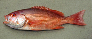
Photography. Coloration can be important for identification of many fish species. Because bright colors fade rapidly after death, photographs should be taken as soon as possible after collection, and prior to fixation if possible. Lateral photographs of the left side are most useful for taxonomic purposes. For some specimens, closeup views of the head are also useful. Techniques for illuminating and positioning of specimens and their fins are reviewed in Randall (1961), Emery and Winterbottom (1980) and Flescher (1983).
4. Fixing and preserving specimens
Methods of fixing and preserving of fishes are extensively discussed in a 1979 publication by The American Society of Ichthyologists and Herpetologists, "A Report on Current Supplies and Practices Used in Curation of Ichthyological Collections." It is available on-line at: http://www.asih.org/coms/ihcc/news/1979.pdf. Widely accepted practices are summarized below.
Fixation. Adult fish specimens are fixed in 10% buffered formalin while larvae are generally fixed in 5% buffered formalin. For large specimens (> 150-200 mm standard length) the right side of the abdominal cavity should be slit to facilitate entrance of fixative. Larger specimens should be injected throughout the body including the lateral musculature. Specimens should remain in formalin for 7 to 10 days or longer for very large specimens.
Preservation. Fishes are most often preserved long-term in 70% ethanol, but several collections use 50% isopropanol. Prior to transfer to alcohol for preservation, formalin should be washed out in one or more changes of water. Because water leaches from tissues, final alcohol percentage may need to be adjusted to the desired level. For long-term storage, the volume of specimens should generally not exceed 2/3 the total volume of specimens and alcohol. Larval fishes are generally stored long-term in 5% buffered formalin to avoid shrinkage and distortion caused by alcohols.
Large specimens are logistically difficult to handle and store. For well-known species, a photographic voucher is often sufficient for positive identification. For sharks, the head and/or jaws are taxonomically informative and may be retained in lieu of the entire specimen.
DNA samples. Several tissues are suitable for DNA extraction from fishes. These include the following. 1) Musculature: remove one or more cubes (5-7 mm) of lateral muscle from the right side of the specimen. 2) Fin clips: remove fin rays and membrane for the right pectoral or pelvic fin. 3) Gill tissue: remove one or more gill arches with attached filaments from the right side. 4) Eye: remove the right eye from extremely small specimens such as larvae. For species with small body size, entire specimens can be placed in preservative in lieu of subsampling. This should be avoided unless a series of conspecifics are available for fixation in formalin for standard morphological analysis.
Tissues samples for DNA extraction should be frozen or preserved in 95% ethanol. Large pieces of tissue should be cut into small pieces (< 5-7 mm) to permit adequate fluid penetration. Tissue samples should be changed to fresh 95% ethanol after a few hours and samples should be stored in cool place, preferably in a freezer.
References and Links
Curation Newletter, http://www.asih.org/coms/ihcc/news/newsletter.html. An occasional publication of the American Society of Ichthyologists and Herpetologists reporting issues and advances in the maintenance of natural history collections.
Emery, A.R. and R. Winterbottom. 1980. A technique for fish specimen photography in the field. Canadian Journal of Zoology 58:2158-2162.
Eschmeyer, W. N. 1998. Catalog of Fishes. Calif. Acad. Sci., San Francisco. Regularly updated on-line version available at: http://www.calacademy.org/research/ichthyology/catalog. Summarizes the taxonomy and nomenclature of fishes.
FishBase, http://www.fishbase.org. On-line clearing house for information on fishes.
Fishnet, http://speciesanalyst.net/fishnet. A distributed information system that permits simultaneous searching for fish specimens in several museum collections.
Flescher, D.D. 1983. Fish photography. Fisheries 8:2-6.
Guidelines for the Use of Fishes in Research, http://www.fisheries.org/html/Public_Affairs/ Sound_Science/Guidelines2004.shtml. An overview of best practices for capture and handling of fishes published in 2004 by the American Fisheries Society.
Hubbs, C.L. and K.F. Lagler. 1958. Fishes of the Great Lakes Region. University of Michigan Press, Ann Arbor, Michigan. Provides standard techniques for morphological study of fishes.
Nelson, J.S. 1994. Fishes of the World. 3rd edition, Wiley & Sons, New York. Summarizes the current taxonomy of fishes.
Randall, J.E. 1961. A technique for fish photography. Copeia 1961:241-242.
Report on Current Supplies and Practices Used in Curation of Ichthyological Collections, http://www.asih.org/coms/ihcc/news/1979.pdf. An overview of best practices for preserving fish specimens published in 1979 by the American Society of Ichthyologists and Herpetologists.
|
|
||||